MRI vs. CT Scan: Which One Does Your Medical Condition Need ?
Medical imaging has transformed how doctors diagnose and treat diseases. Gone are the days of exploratory surgery or guesswork. Today, two technologies stand out: Computed Tomography (CT) and Magnetic Resonance Imaging (MRI). While both create detailed cross-sectional images of your body, they aren’t interchangeable. Choosing the right scan can mean the difference between a quick diagnosis and a missed finding.
Before Your Imaging Examination
What Are MRI Scans and CT Scans?
Both MRI and CT scans provide detailed images of the body's internal structures, helping physicians diagnose a wide range of medical conditions. However, they use different technologies and are best suited for different clinical situations.
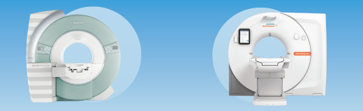
A CT scan uses X-rays and computer processing to create cross-sectional images of the body. It is fast, widely available, and particularly effective for evaluating bones, lungs, internal bleeding, and emergency conditions.
An MRI scan uses powerful magnets and radio waves to generate highly detailed images of soft tissues without exposing patients to ionizing radiation. MRI is especially valuable for assessing the brain, spinal cord, joints, muscles, ligaments, and certain types of tumors.
MRI does not use radiation, making it a preferred imaging option for many conditions requiring repeated follow-up examinations. CT scans, however, can acquire detailed images within seconds, making them indispensable in emergencies where rapid diagnosis can save lives.
During Your Imaging Assessment
MRI vs. CT Scan: The Quick Difference
Healthcare providers may recommend either a MRI scan or CT depending on the suspected condition, symptoms, body region being examined, and urgency of diagnosis.
- MRI (Magnetic Resonance Imaging): Uses powerful magnets and radio waves. It provides superior soft-tissue detail without radiation and is ideal for imaging the brain, spine, joints, and many tumors.
- CT Scan (Computed Tomography): Uses X-rays. It is fast, widely available, and excellent for evaluating bones, lungs, fractures, and bleeding.
- Neurology & Neurosurgery: CT is preferred for acute stroke, head trauma, skull fractures, and brain bleeds. MRI is preferred for brain tumors, multiple sclerosis, epilepsy, spinal cord disorders, and early stroke detection.
- Orthopedics & Sports Medicine: CT helps evaluate complex fractures and bone deformities. MRI is the gold standard for ACL tears, meniscal injuries, rotator cuff tears, cartilage damage, and bone marrow abnormalities.
- Oncology (Cancer Care): CT is commonly used for tumor detection, cancer staging, and monitoring treatment response. MRI provides superior evaluation of brain tumors, liver lesions, prostate cancer, rectal cancer, and soft tissue sarcomas.
- Cardiology: CT is useful for coronary calcium scoring, coronary CT angiography, and aortic aneurysm evaluation. MRI helps assess cardiomyopathies, myocarditis, congenital heart disease, and myocardial viability.
- Pulmonology: CT is the preferred imaging method for lung cancer screening, pulmonary embolism, interstitial lung disease, and pneumonia. MRI has limited use for mediastinal and chest wall tumors.
- Gastroenterology: CT is valuable for appendicitis, pancreatitis, bowel obstruction, and abdominal trauma. MRI excels in liver lesion characterization, MRCP imaging, and inflammatory bowel disease assessment.
- Urology: CT remains the gold standard for kidney and ureteric stones. MRI is widely used for prostate cancer assessment and characterization of renal tumors.
- Obstetrics & Gynecology: MRI provides detailed assessment of endometriosis, adenomyosis, uterine anomalies, ovarian masses, and fetal abnormalities while avoiding radiation exposure.
- ENT: CT is preferred for sinus disease, temporal bone disorders, and facial fractures. MRI is ideal for acoustic neuromas, skull base tumors, neck masses, and soft tissue lesions.
- Emergency & Critical Care: CT is the first-line imaging tool for trauma, internal bleeding, acute chest pain, and emergency stroke evaluation because of its speed. MRI is reserved for selected neurological emergencies.
- Vascular Surgery: CT angiography is commonly used for aortic aneurysms and peripheral arterial disease. MR angiography helps evaluate vascular malformations and venous disorders without radiation.
After Your Imaging Examination
Why These Scans Matter
MRI and CT scans provide healthcare professionals with essential information that can improve diagnostic accuracy, guide treatment decisions, and monitor disease progression.
The Importance of Choosing the Right Imaging Test
- Accurate Diagnosis: Selecting the appropriate imaging modality improves the likelihood of identifying the correct condition.
- Early Detection: Advanced imaging can reveal disease before symptoms become severe.
- Treatment Planning: Imaging findings help physicians determine the most effective treatment strategy.
- Monitoring Progress: Repeat imaging allows healthcare providers to evaluate treatment response and disease progression.
- Improved Patient Outcomes: Early and accurate diagnosis often leads to better outcomes and reduced complications.
Understanding Your Results
MRI and CT: Complementary Tools in Modern Medicine
MRI and CT are complementary tools rather than competing technologies. Each has unique strengths that make it valuable in specific clinical situations.
CT is typically preferred when speed, bone detail, lung imaging, or emergency evaluation is required. MRI is often the best choice when superior soft-tissue detail, neurological assessment, joint evaluation, or radiation-free imaging is needed.
Emerging technologies such as Artificial Intelligence (AI), functional MRI, synthetic MRI, dual-energy CT, and radiomics are making medical imaging faster, safer, and more precise. These innovations continue to improve diagnostic accuracy and support personalized patient care.
The ultimate goal remains the same: providing the right imaging test at the right time to achieve better patient outcomes and more effective treatment decisions.
This article is for educational purposes only and should not replace professional medical advice. Always consult a qualified healthcare professional for diagnosis and treatment recommendations.